A Bird's Eye View of
Brain Bank Functioning
The complete journey — from a family's call to a researcher's stained slide.
The Brain Donation Process
Receiving the Call
Brain Bank staff receives calls from bereaved families, and provides assistance for arranging transport to the Mortuary for the postmortem.
- Copy of Death certificate from a Registered Medical Practitioner
- Medical records of patient
- Brain donation card if previously registered
- Photo ID proof of the deceased and proof of relationship of relative signing consent
In the Mortuary
Relatives are explained about the entire procedure. Documents provided by the family will be scrutinized. If documents are in order, signature of first degree relatives on Informed consent form is obtained.
- Postmortem procedure will take 1–1½ hours
- The body will be handed back with due respect. Care is taken to ensure there is no disfigurement.
After Postmortem
Following documents should be collected from the Mortuary:
- Acknowledgement form of brain/spinal cord donation from Brain Bank
- Copy of informed consent signed
- Death certificate (original)
Following Donation
You receive the following from the Brain Bank:
- Thanks giving letter for donation along with Certificate of donation
- Final postmortem report (within 3 months)
- Annual update on information about Brain Bank activities
From Postmortem Table to Research Slide
Each step in the exact order it happens — the science behind every donated brain.
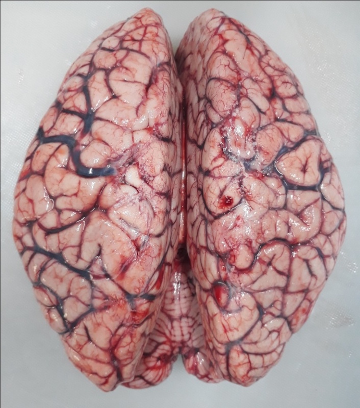
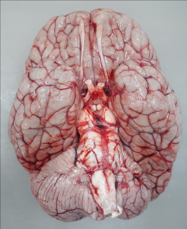
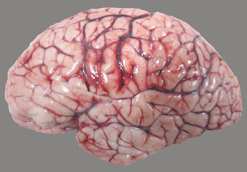
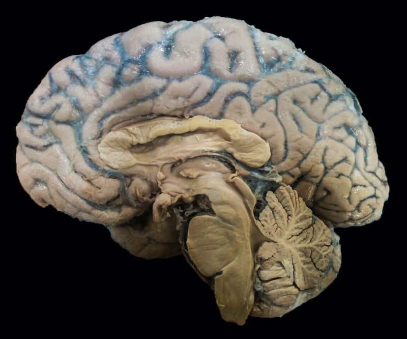
Step 1: The brain is examined for any abnormalities
Fresh brain extracted following postmortem
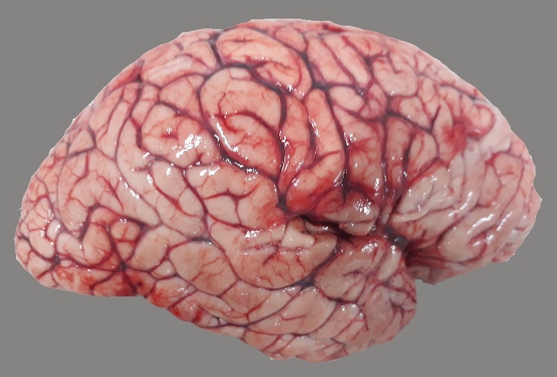
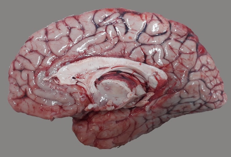
Step 2: The brain is bisected into 2 halves
One half of the brain will be sliced for freezing at −86°C. The other half will be fixed in 10% neutral buffered formalin for examination of pathology under microscope.
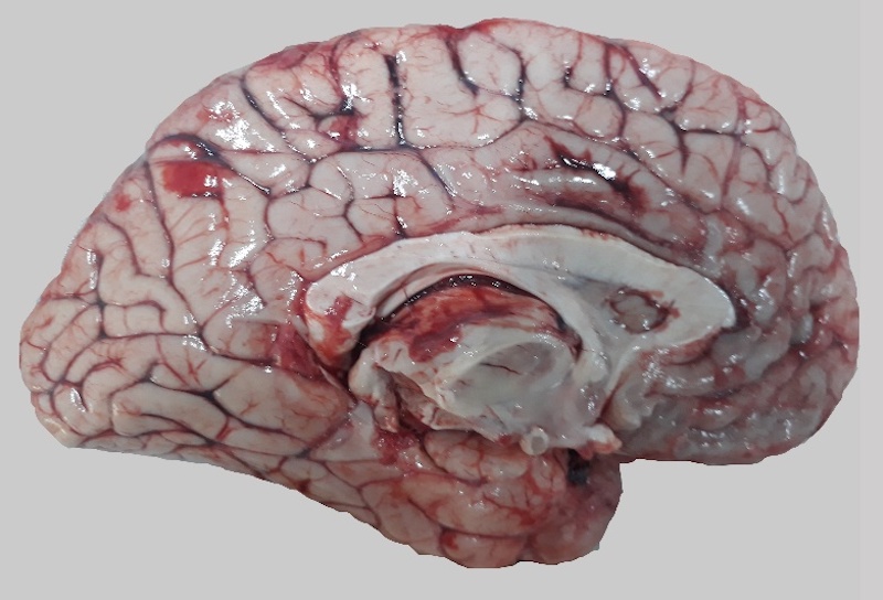
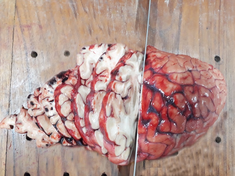
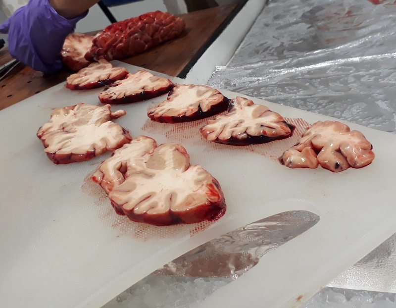
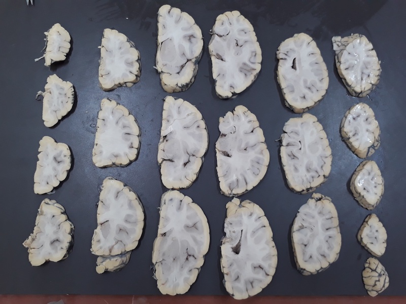
Slicing the cerebral hemisphere
Coronal sections: One half of the brain is being sliced from front to back (like bread loaf) serially. The slices are arranged serially on a dissection board, carefully examined and photographed.
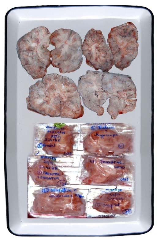
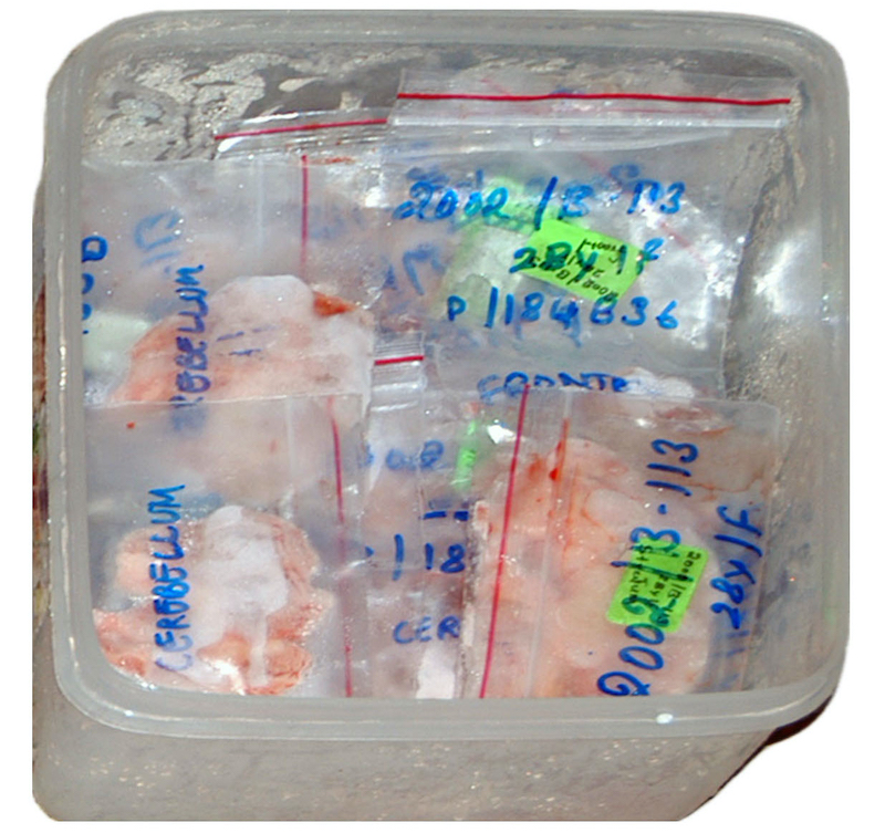
Storing of fresh brain tissue
The brain slices are placed carefully in Ziplock cover, each labelled with case number, and anatomical site. The ziplock bags are placed in a box and stored in a deep freezer that keeps the tissue at −86°C, awaiting use by researchers.
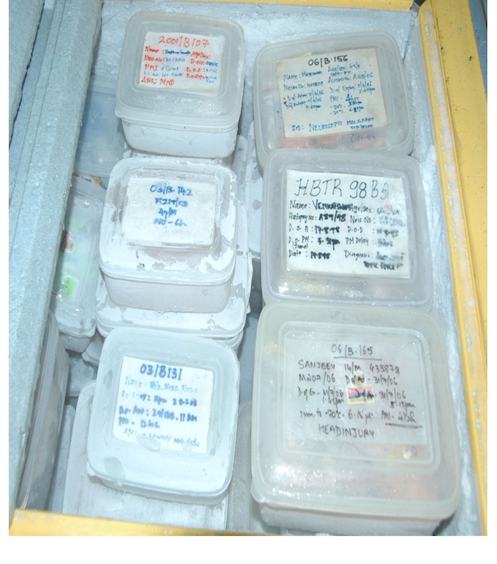
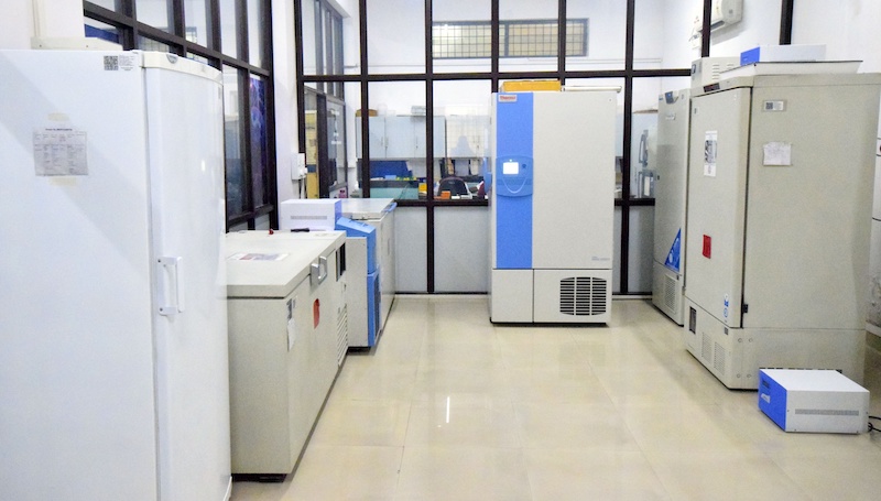
Freezer Room
The Brain Bank's freezer rooms. All the freezers are connected to a temperature alarm system that is monitored 24/7.
THE other half…
The other half of the brain is immersed in buffered neutral formalin for a minimum of 3–4 weeks. This half of the brain is examined under a microscope by a neuropathologist to ascertain if brain tissue is normal or has any disease process, and a final diagnosis based on the examination is provided.
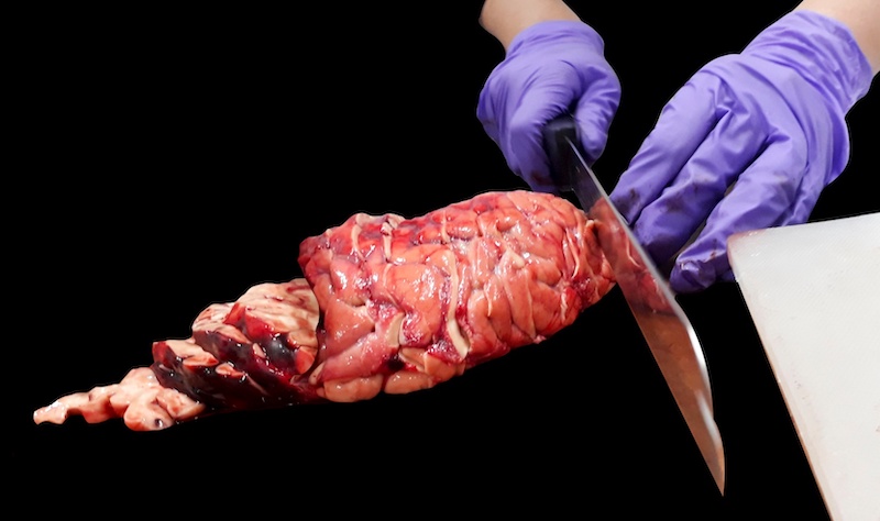
THE Gross Examination
To examine the brain, it is first sliced and the interior of the slices are examined.
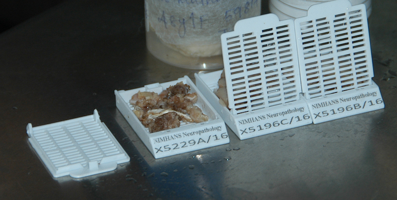
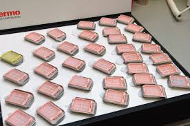
Sampling the brain
The neuropathologist makes 20–25 blocks of tissue (~2×2 cm sized) from different anatomical areas to be studied microscopically. These are then placed into labelled cassettes to "process" the tissues.
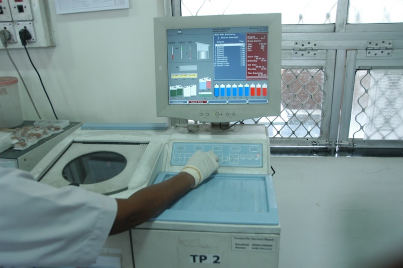
Processing the tissue
These tissue cassettes are then put into a "tissue processor" and then "embedded" in molten paraffin wax to produce a "block". The wax will harden the tissue so that it can be sliced into extremely thin sections.
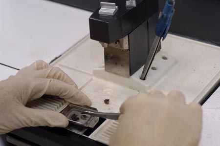
Embedding the tissue
A bit of brain tissue is put into a mold and embedded by paraffin wax. To the left is the freezing plate where the blocks are hardening.
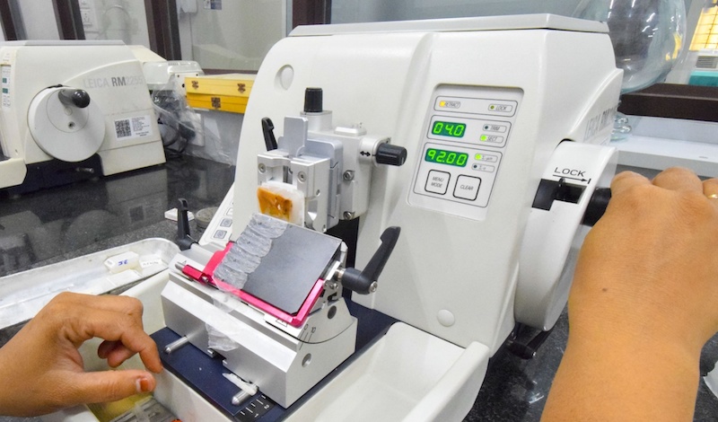
Sectioning of the tissue
Thin sections are cut from the wax blocks on a microtome.
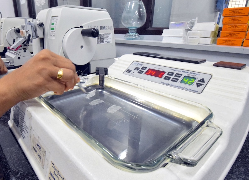
Sectioning of the brain
The tissue sections are spread out on a heated water bath and are collected onto glass slides. These sections will be stained with various dyes to show the microscopic structure of the brain tissue and assess changes caused by various diseases, if any.
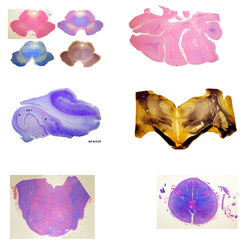
Set of stained slides from embedded brain tissue
A set of stained sections taken from the blocks of brain tissue, showing grey matter, white matter tracts and other cellular details of the tissue.
Every Brain Tells a Story
The Brain Bank Network preserves these stories — for the researchers of today and the treatments of tomorrow. Consider pledging your brain or supporting our mission.